Home | Fields of Research | Members


Rotating-Frame NMR Microscopy

with a Toroid Cavity Detector


The Inside Information

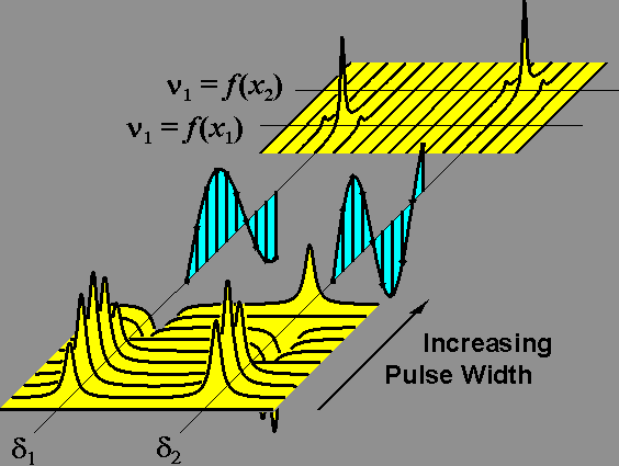
Figure: One dimensional rotating-frame images are obtained from a set of NMR spectra collected with incrementally increasing pulse width (lower yellow plot). The amplitude modulated spectra, where the modulation frequency ist the nutation frequency (cyan plot), are converted (by a second real Fourier transformation) into a two-dimensional plot revealing spectral information in the one and spatial information in the other dimension (upper yellow plot). If the dependency of nutation frequencies on the spatial dimension is known, the locations of NMR-active nuclei can be calculated.

  • Rotating-Frame Imaging (RFI) is one of the many Magnetic Resonance Imaging (MRI) methods that can be used to obtain images without disecting the sample.

  • The main feature of RFI is a radiofrequency-field gradient (B1). This gradient is used instead of a static field gradient (B0) to encode spatial information.

  •  


    (The nutation of magnetization instead of the Larmor precession is used as the imaging parameter)


  • Advantages:
    • Chemical Shift information is not sacrifized for spatial information,
    • Spatial resolution is less dependent on the transversal relaxation time (T2).


Additional information can be obtained from the further reading page or by e-mail request.


Back to Toroid Cavity NMR Spectroscopy and Imaging


K. Woelk, October 22, 1998